Cell Organelles: Specialized Parts Working Together
Students interpret and create a scale-based cell model to explain how organelles perform specialized functions that help a cell survive.

Illustrations are auto-generated and may be placeholders. They can be refreshed to match the narration.
Inside a Cell
A cell is the smallest unit that can carry out all processes needed for life. The cell membrane surrounds the cell and controls what enters and leaves. Inside, cytoplasm holds specialized structures called organelles. Each organelle performs particular jobs, but the entire cell functions as one system. Most organelles are too small to see without a microscope. In the 1600s, improved lenses allowed Robert Hooke to observe boxlike compartments in cork and Antonie van Leeuwenhoek to view living microscopic organisms. Later advances in light and electron microscopy revealed more cell structures, changing scientific explanations of life. For example, electron microscopes helped scientists see the folded inner membrane of a mitochondrion. This structure supports the organelle’s role in releasing usable energy from food. Cell diagrams and models make these tiny, three-dimensional structures easier to study.

Organelles and Their Functions
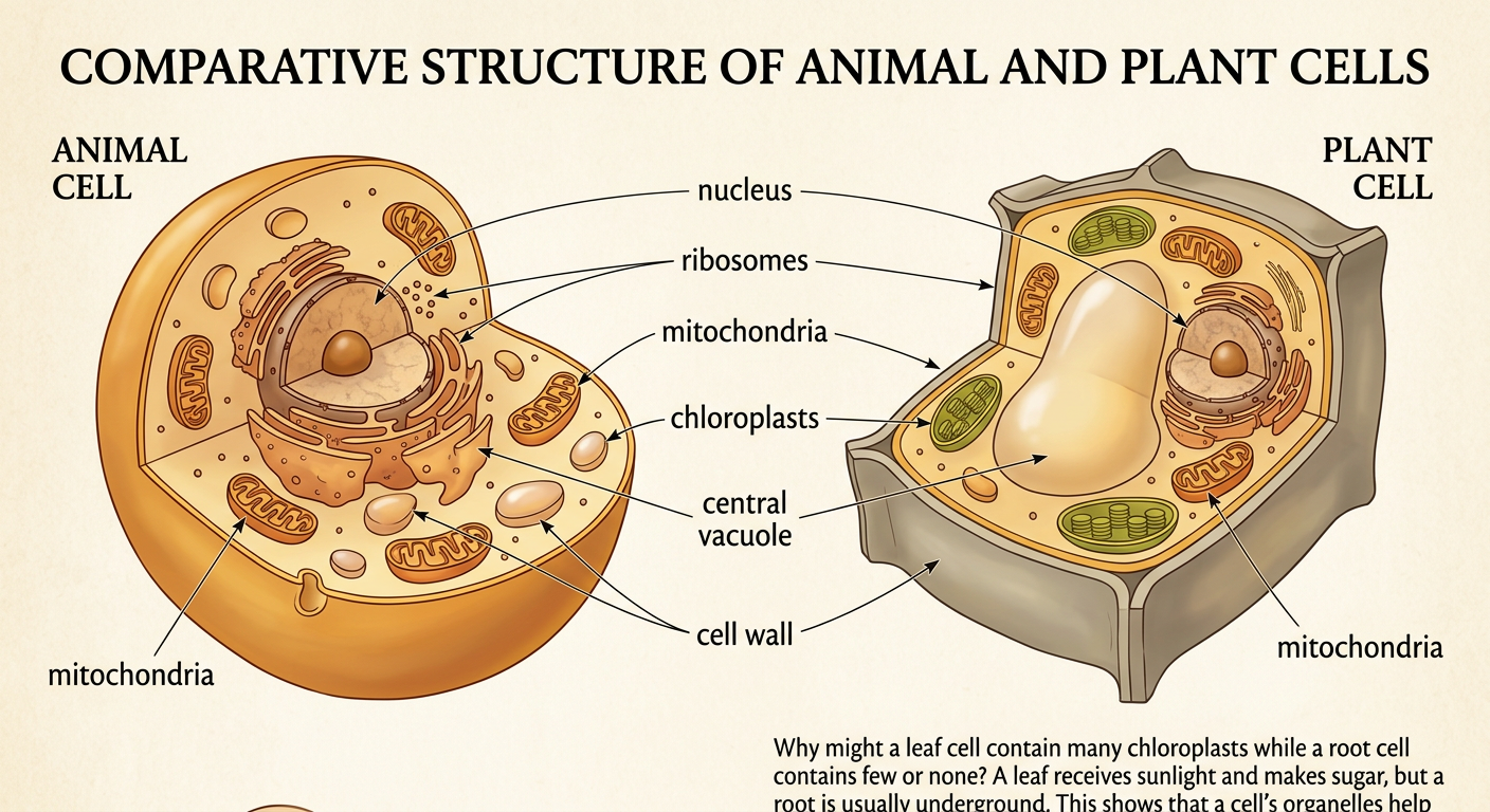
Organelles have structures suited to their functions. The nucleus contains most of the cell’s DNA, which provides instructions for cell activities. Ribosomes build proteins. The rough endoplasmic reticulum helps fold and transport proteins, while the Golgi apparatus modifies, sorts, and packages them. Mitochondria release usable energy from food through cellular respiration. Lysosomes contain substances that break down worn-out cell parts and some large molecules. Vacuoles store water and other materials. Plant cells also have chloroplasts, where photosynthesis captures light energy, a large central vacuole that helps maintain shape, and a rigid cell wall outside the membrane. For example, a leaf cell contains many chloroplasts because it receives sunlight and produces sugars. A root cell usually lacks chloroplasts because it is underground. The organelles present in a cell help match that cell to its job.
How Cell Parts Work Together
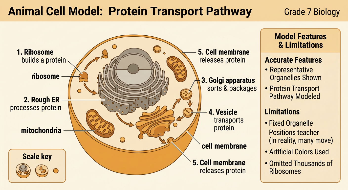
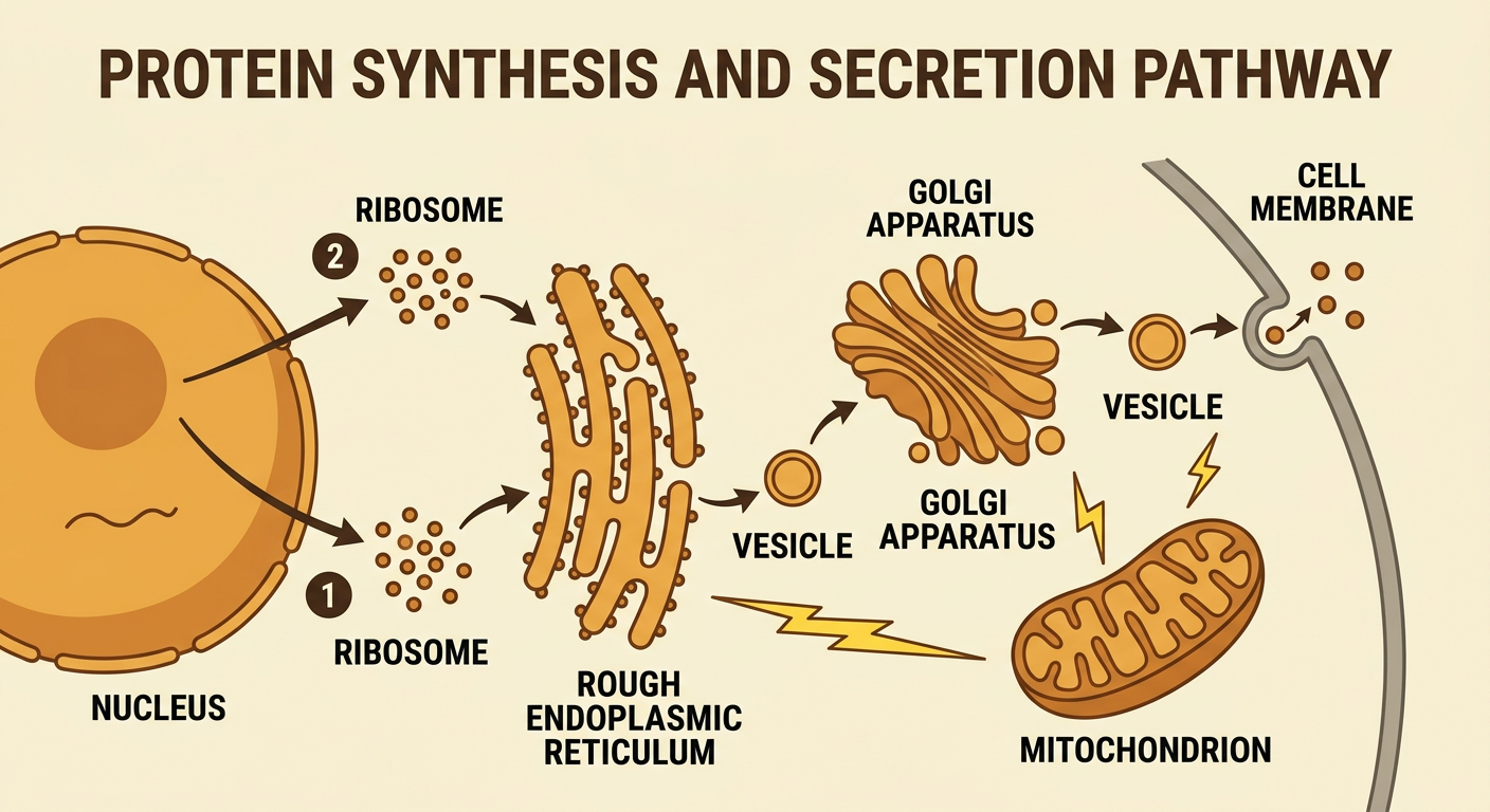
A cell survives because its parts interact rather than work alone. Consider a cell making and releasing a protein. DNA in the nucleus contains the instructions. A ribosome reads a copied message from the DNA and joins amino acids to build the protein. If the protein will leave the cell, the rough endoplasmic reticulum helps process and transport it. A transport vesicle carries it to the Golgi apparatus, where it is modified and packaged. Another vesicle moves the finished protein to the cell membrane, which releases it outside the cell. Mitochondria provide usable energy for many steps in this process. For example, cells in a salivary gland make and release the protein enzyme amylase, which helps break down starch. If one part of this pathway fails, protein production or delivery may be reduced, affecting the cell and the organism.
Reading a Cell Diagram
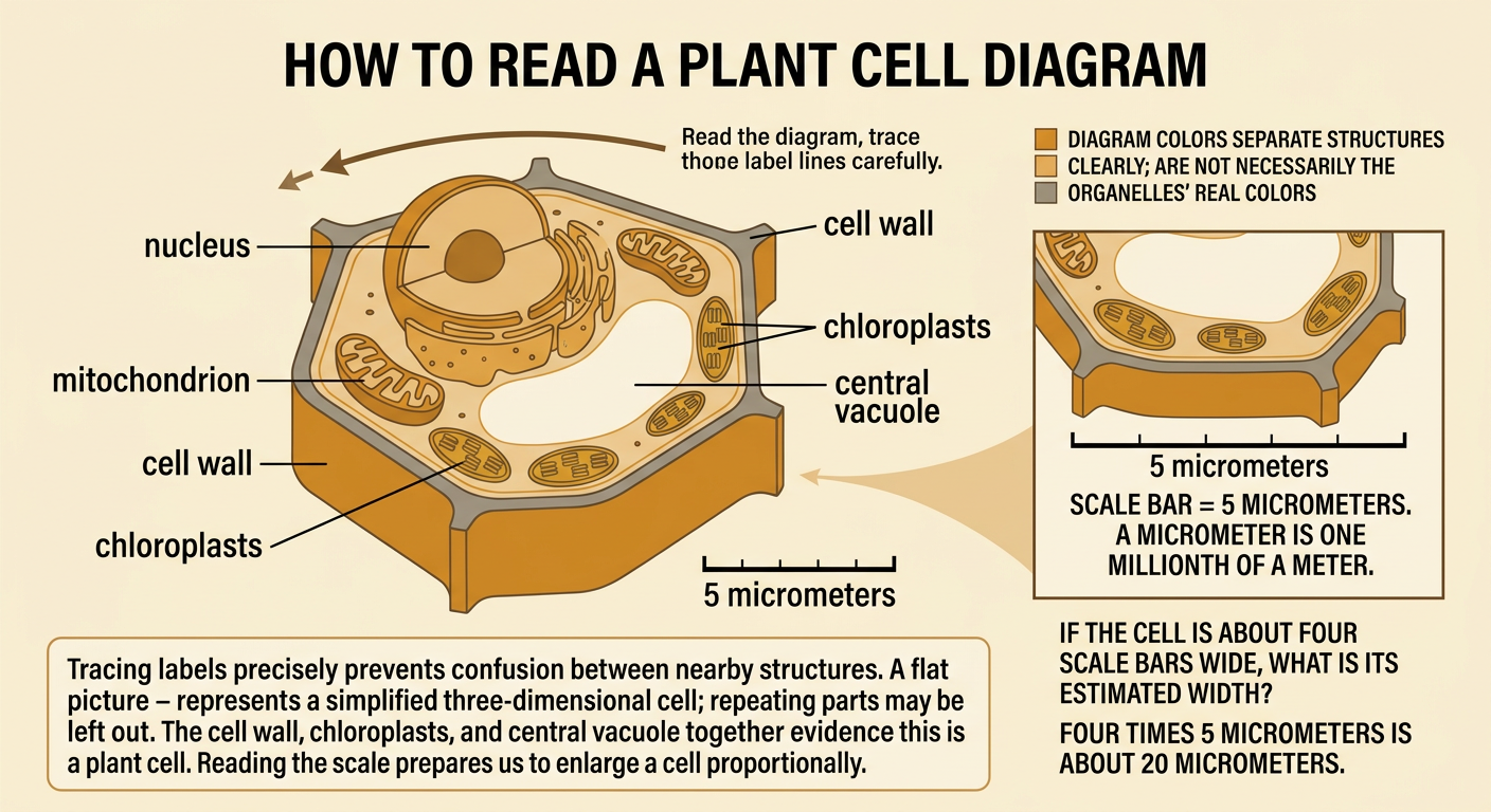
To interpret a cell diagram, first read its title, key, labels, arrows, and scale information. Trace each label line carefully so you do not confuse nearby structures. Then connect visual evidence with written information about function. A nucleus is usually drawn as a large structure surrounded by a nuclear envelope, while mitochondria are often shown with folded inner membranes. Colors in textbook diagrams are usually chosen to separate structures; they are not necessarily the organelles’ actual colors. Also remember that a flat diagram represents a three-dimensional cell and may simplify shapes or leave out repeated structures. For example, if a diagram shows a rigid outer boundary, chloroplasts, and one large central vacuole, evidence supports identifying it as a plant cell. If a scale bar represents 5 micrometers, compare the bar with the cell’s width to estimate the cell’s actual size.
Building a Scale-Based Cell Model
A scale model enlarges a cell while keeping measurements proportional. Suppose a cell is 20 micrometers wide and you choose a scale of 1 micrometer to 1 centimeter. The model must be 20 centimeters wide. If the actual nucleus is 6 micrometers wide, its model width is 6 centimeters. The proportional relationship can be written as model size in centimeters equals actual size in micrometers times 1 centimeter per micrometer. Use the same scale for every measured structure, and record calculations in a table before building. Position each organelle according to reliable cell images, but do not crowd every organelle into one flat layer because real cells are three-dimensional. For example, a mitochondrion measuring 2 micrometers long would be 2 centimeters long in this model. Include a key, a scale statement, labels, and arrows showing important interactions among organelles.

Explain the Model
A useful model must be explained with evidence, not simply displayed. Begin by identifying the type of cell and the scale used. Describe how each represented organelle contributes to the cell’s overall survival, and use arrows or a sequence to show interactions. For example, explain that a ribosome builds a protein, the rough endoplasmic reticulum helps process it, the Golgi apparatus sorts and packages it, and a vesicle carries it to the cell membrane. Refer directly to model features, such as labels, relative sizes, and arrow paths. Also state the model’s limitations. A classroom model may show organelles as fixed objects even though many structures move and change. It may use artificial colors and omit thousands of ribosomes. Finish by explaining cause and effect: if mitochondria cannot release enough usable energy, many energy-requiring cell activities slow down, so the cell may not maintain stable conditions.