DNA Copying Changes: Types and Causes of Gene Mutations
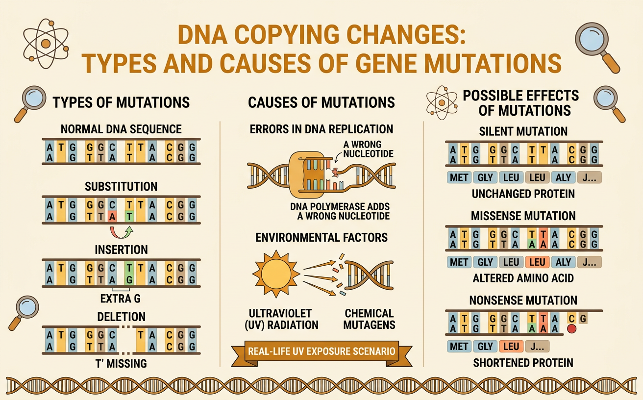
Students compare DNA sequences to identify substitutions, insertions, and deletions, examine causes and possible effects of mutations, and apply their understanding to a real-life UV exposure scenario.
Illustrations are auto-generated and may be placeholders. They can be refreshed to match the narration.
What Is a Gene Mutation?
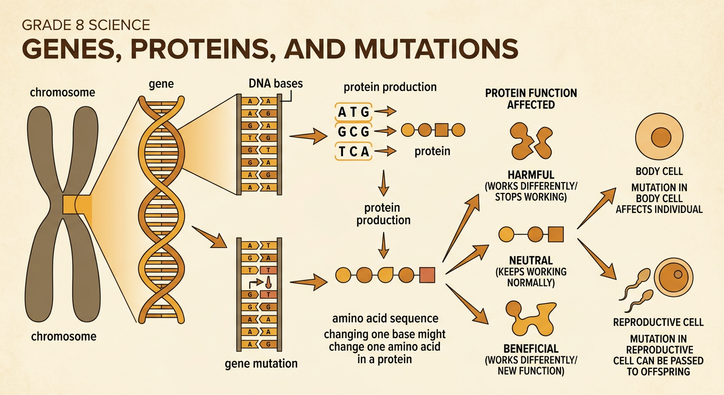
A gene mutation is a change in the order or number of DNA bases within a gene. Genes are sections of DNA located on chromosomes. Many genes contain instructions for assembling proteins, which help build structures and control cell processes. During protein production, the cell reads DNA information in groups of three bases. A mutation may change the instructions, but its effect depends on where the change occurs. For example, changing one DNA base might produce a different amino acid in a protein. The altered protein could work differently, stop working, or continue working normally. Therefore, mutations can be harmful, beneficial, or neutral. Mutations in body cells affect only the individual, while mutations in reproductive cells can sometimes be passed to offspring.
Comparing Original and Mutated DNA Sequences
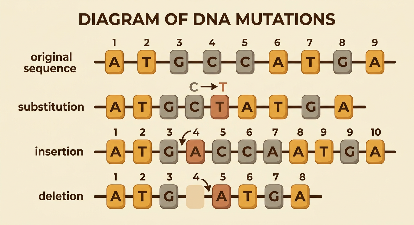
DNA sequences can be compared by lining them up and checking each base from left to right. Consider the original sequence A T G C C A T G A. A mutated sequence, A T G T C A T G A, has the same length, but C at position 4 has been replaced by T. This is one difference out of nine bases. Another mutated sequence, A T G A C C A T G A, contains an extra A after position 3 and is ten bases long. A third sequence, A T G C A T G A, is missing one C and is eight bases long. Using aligned rows, position numbers, and color-coded differences helps connect written descriptions with visual evidence. Sequence length and the location of mismatched bases provide evidence for identifying the type of mutation.
Substitutions, Insertions, and Deletions
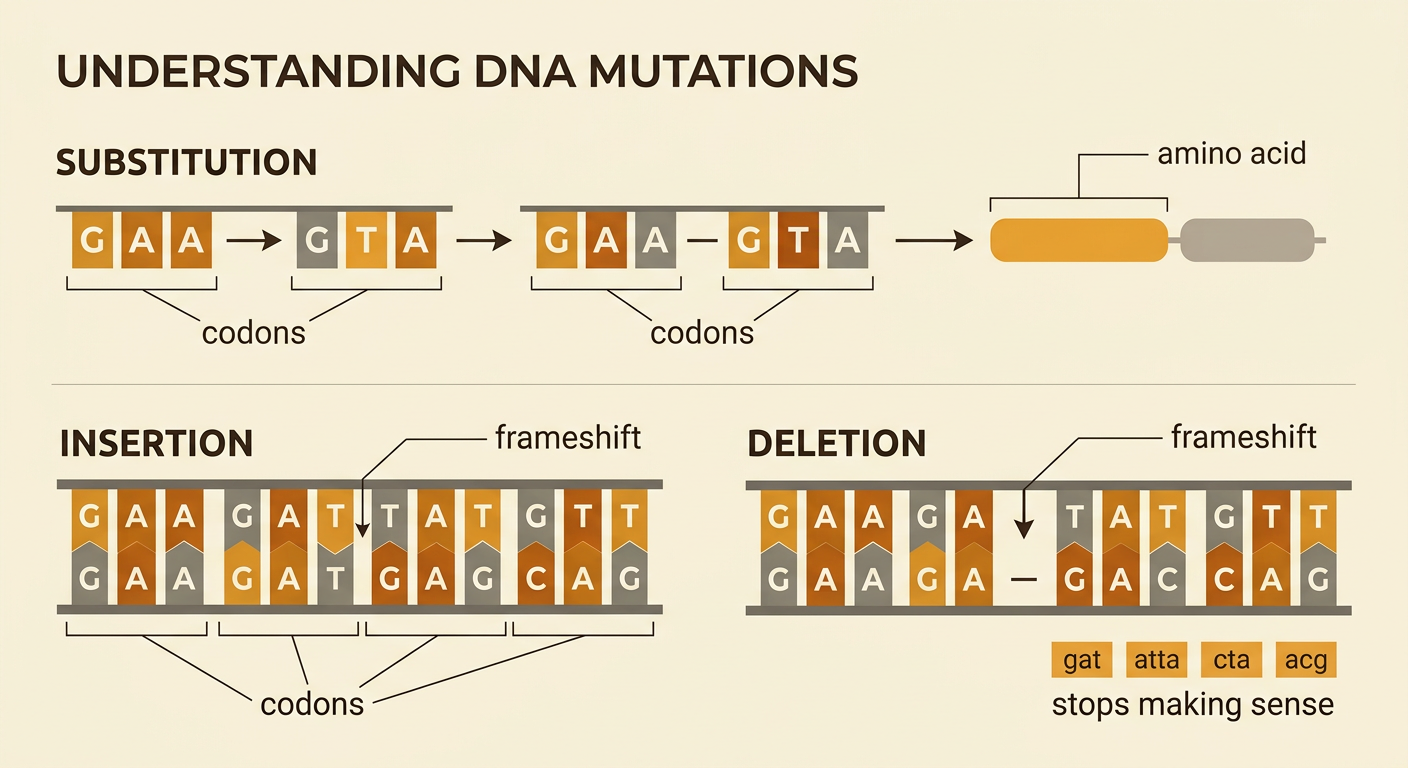
A substitution occurs when one DNA base is replaced by another. For example, the coding DNA triplet GAA may change to GTA, which can specify a different amino acid. An insertion adds one or more bases, while a deletion removes one or more bases. Cells read a protein-coding sequence in three-base units called codons. If one base is inserted or deleted, the grouping of every codon after that point usually shifts. This is called a frameshift and can greatly change the protein. For example, CAT-GAA-TTC could become CCA-TGA-ATT after one base is inserted near the beginning. A substitution may be neutral if the new codon specifies the same amino acid. Insertions and deletions of exactly three bases may add or remove one amino acid without shifting the remaining reading frame.
Causes: Copying Errors, Radiation, and Chemicals
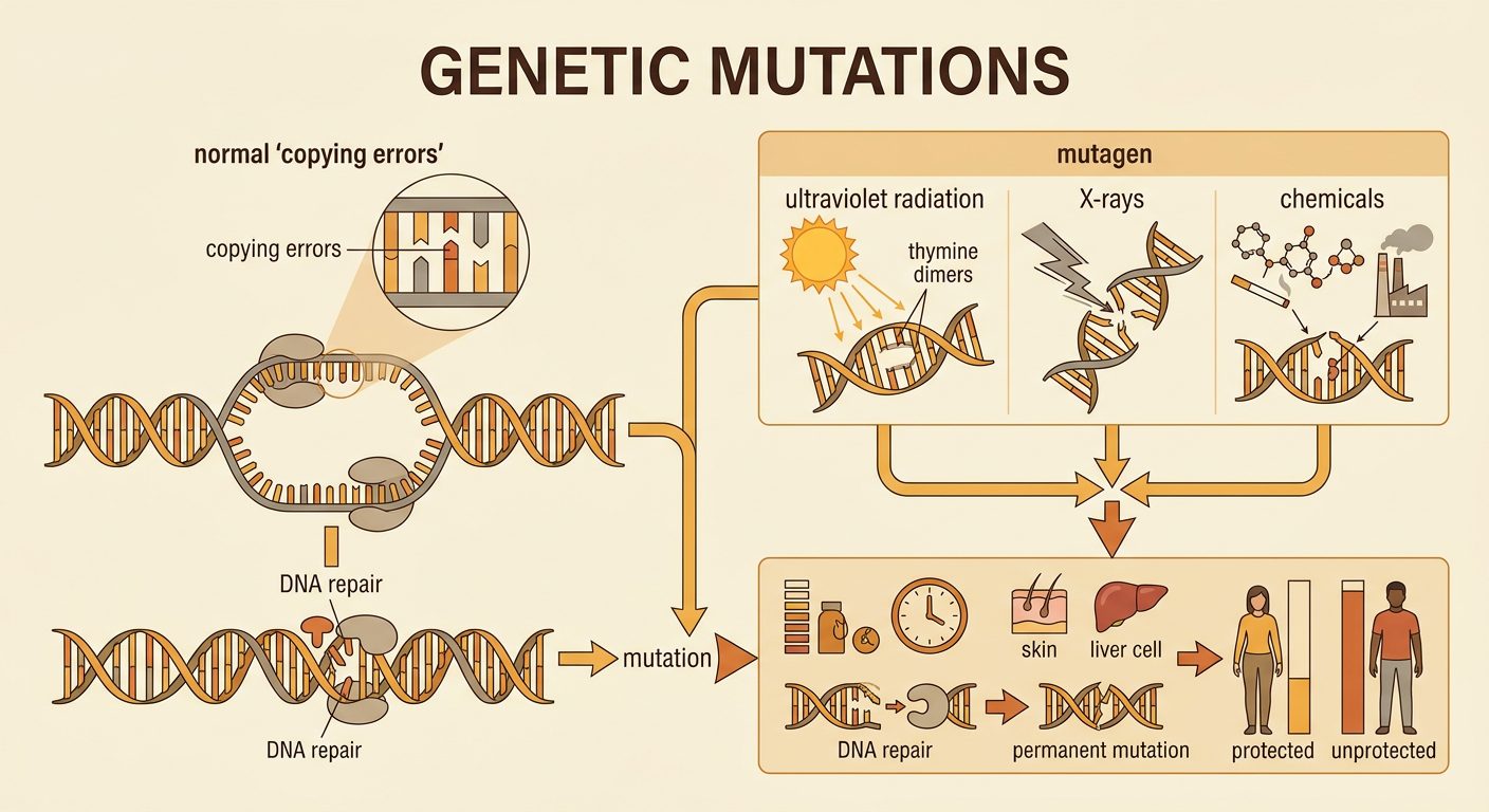
Mutations can occur spontaneously or after exposure to a mutagen, an agent that increases the chance of DNA damage. Before a cell divides, enzymes copy its DNA. Most errors are corrected by proofreading and DNA repair systems, but an uncorrected error can become a mutation after another round of copying. Radiation can also damage DNA. Ultraviolet radiation from sunlight can link neighboring thymine bases, bending the DNA and interfering with accurate copying. Higher-energy ionizing radiation, such as X-rays, can break DNA strands. Some chemicals in tobacco smoke or industrial pollution can react with DNA bases. Exposure does not guarantee a mutation, and one mutation does not guarantee disease. Risk depends on dose, exposure time, cell type, and the success of repair. A mutation becomes permanent only if the altered DNA remains when the cell copies it.
Real-Life Scenario: UV Exposure and Skin-Cell Mutations
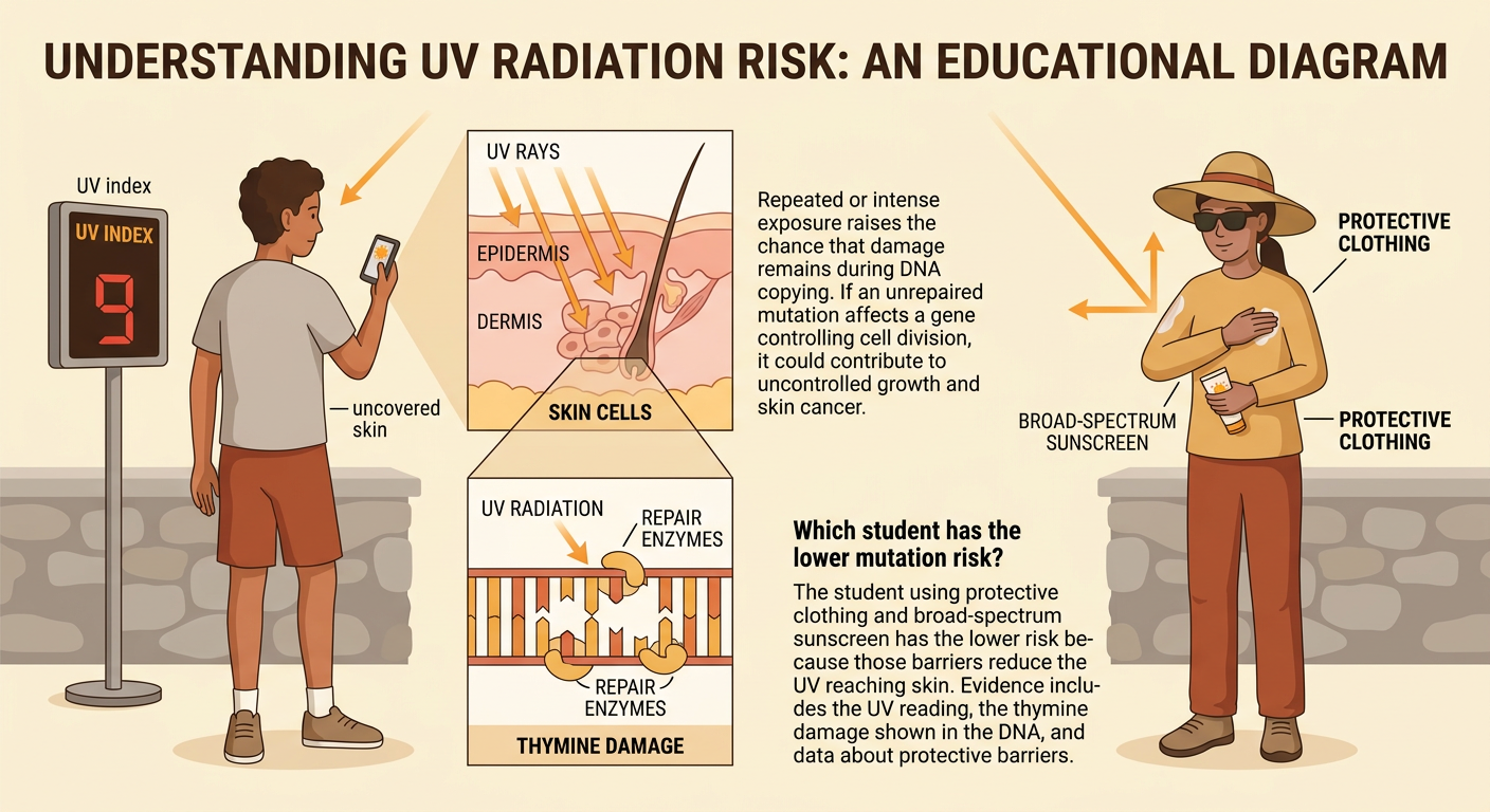
Imagine two students spend the same hour outdoors at midday. One wears protective clothing and broad-spectrum sunscreen, while the other has uncovered skin. A UV meter shows an index of 9, meaning protection is especially important. Ultraviolet radiation can reach skin cells and cause neighboring thymine bases in DNA to bond incorrectly. Repair enzymes often remove this damage, but repeated or intense exposure increases the chance that some damage remains during DNA copying. An unrepaired mutation in a gene controlling cell division could contribute to uncontrolled cell growth and skin cancer. This evidence supports the claim that reducing UV exposure lowers mutation risk, although it cannot remove all risk. Useful evidence includes the UV reading, a diagram of thymine damage, and data showing that protective barriers reduce the amount of UV reaching skin.
Exit Ticket: Identify, Explain, and Predict
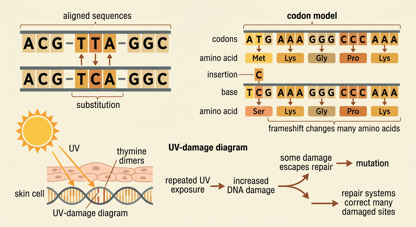
Use sequence evidence and the mutation model to answer three questions. First, compare the original sequence ACG-TTA-GGC with ACG-TCA-GGC. Identify the mutation as a substitution because T was replaced by C and the sequence length stayed the same. Second, explain why adding one base near the beginning of a protein-coding gene may have a larger effect: the insertion can shift later codons and change many amino acids. Third, predict the effect of repeated UV exposure on skin cells. A strong response states that more exposure can create more DNA damage, increasing the chance of unrepaired mutations, while noting that repair systems correct many damaged sites. Support the prediction with at least two sources of evidence, such as the aligned sequences, the codon model, the UV-damage diagram, or exposure data. Do not claim that every mutation is harmful.