Electromagnetic Radiation: Evaluating Risks and Benefits
Students compare how different frequencies of electromagnetic radiation interact with matter and evaluate evidence-based claims about their risks and practical uses.

Illustrations are auto-generated and may be placeholders. They can be refreshed to match the narration.
The Electromagnetic Spectrum
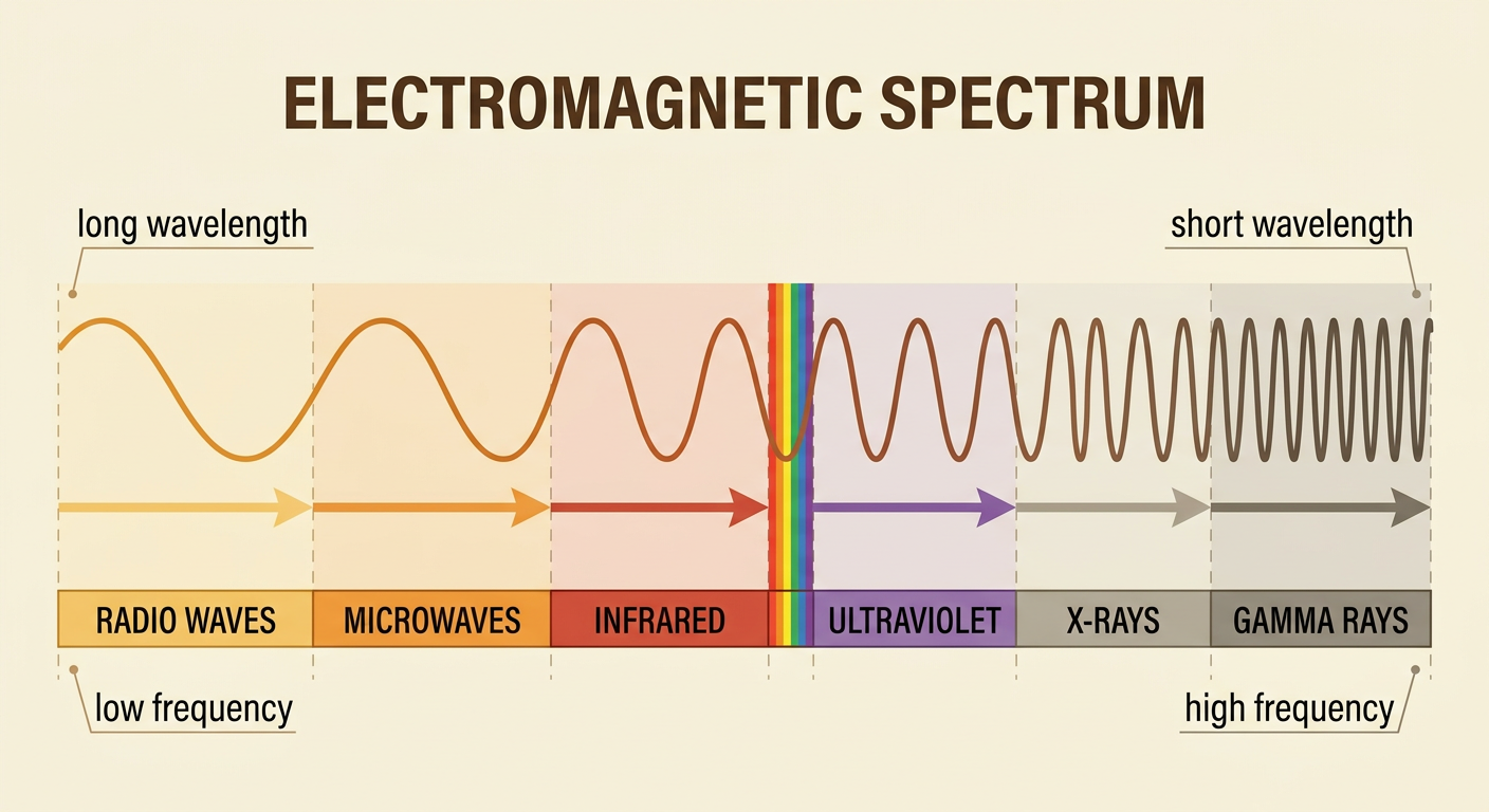
Electromagnetic radiation carries energy through changing electric and magnetic fields. The electromagnetic spectrum arranges this radiation by wavelength or frequency. From lowest to highest frequency, the main regions are radio waves, microwaves, infrared, visible light, ultraviolet, X-rays, and gamma rays. The boundaries between regions are useful categories rather than sharp physical divisions. Frequency increases as wavelength decreases because all electromagnetic waves travel at the same speed in a vacuum. Different parts of the spectrum have practical uses. For example, a radio station transmits low-frequency radio waves that an antenna detects and converts into sound. A hospital may use much higher-frequency X-rays to produce images of bones. Position on the spectrum helps scientists predict how radiation may interact with matter, but exposure amount and the material involved also matter.
Frequency, Energy, and Matter
The energy of one photon of electromagnetic radiation increases with frequency. This relationship is written as energy equals Planck’s constant times frequency. When radiation reaches matter, it may be reflected, transmitted, scattered, or absorbed. Absorption occurs only when the material can take in the photon’s energy through an allowed change. Different frequencies can produce different effects, such as molecular motion, electronic excitation, or ionization. Intensity and exposure time determine how much total energy reaches the material, so frequency alone does not determine risk. For example, skin absorbs infrared radiation from a heat lamp, increasing molecular motion and temperature. Visible light from the lamp may instead be reflected by the skin or absorbed by pigments. Scientists therefore examine both photon energy and total absorbed energy when explaining how radiation affects matter.

Ionizing and Non-Ionizing Radiation
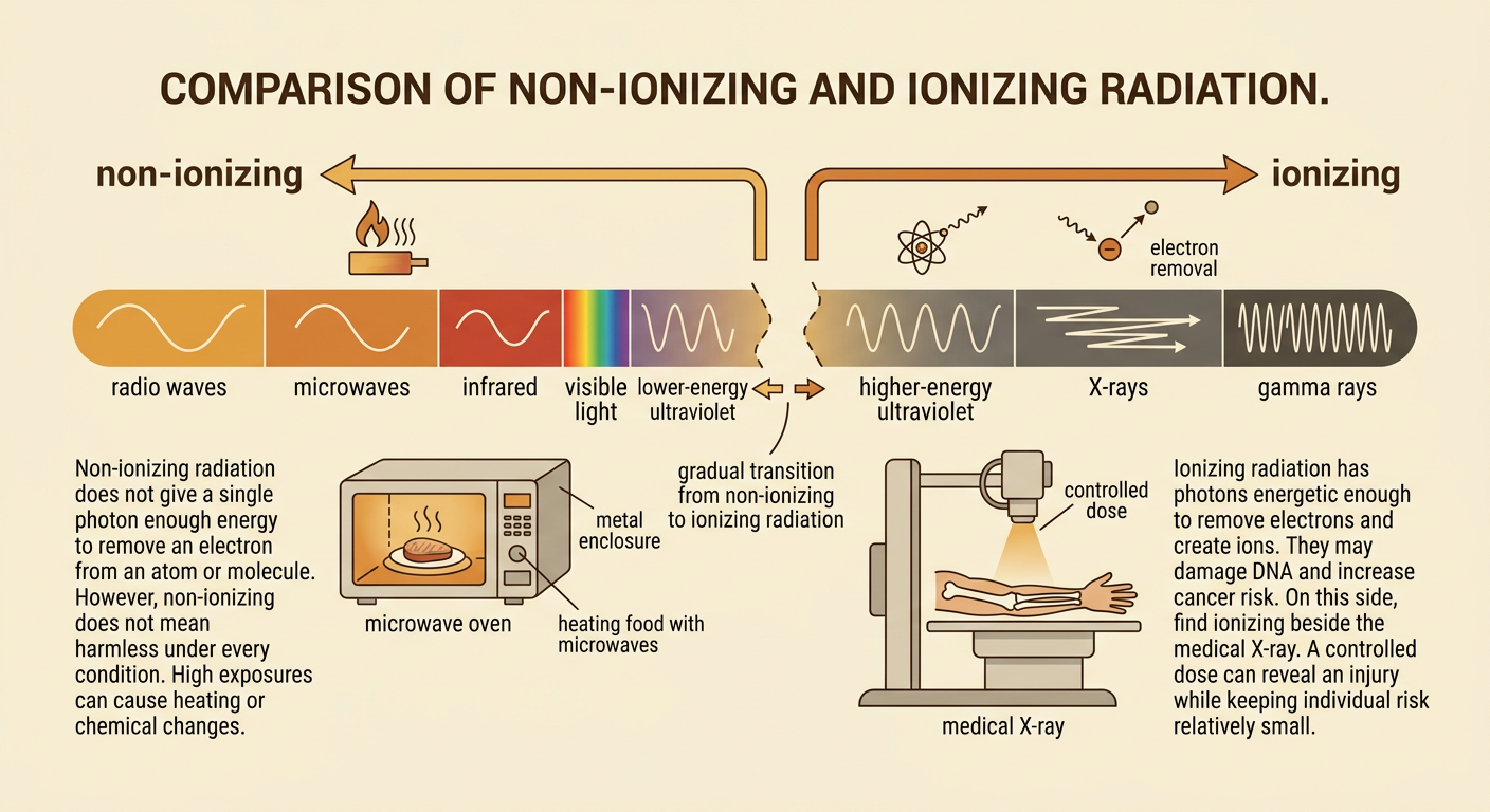
Non-ionizing radiation does not give a single photon enough energy to remove an electron from an atom or molecule. Radio waves, microwaves, infrared, visible light, and lower-energy ultraviolet are generally non-ionizing. They can still cause harm at high exposures, mainly through heating or chemical changes. Ionizing radiation has photons energetic enough to remove electrons and create ions. Higher-energy ultraviolet, X-rays, and gamma rays can ionize matter and may damage DNA, increasing cancer risk. Risk is not determined by category alone; absorbed dose, exposure time, exposed tissue, and shielding are important. For example, a microwave oven uses non-ionizing microwaves to heat food, but its metal enclosure limits exposure outside the oven. A medical X-ray is ionizing, yet a carefully controlled dose can provide valuable diagnostic information with a relatively small individual risk.
Evaluating Scientific Claims
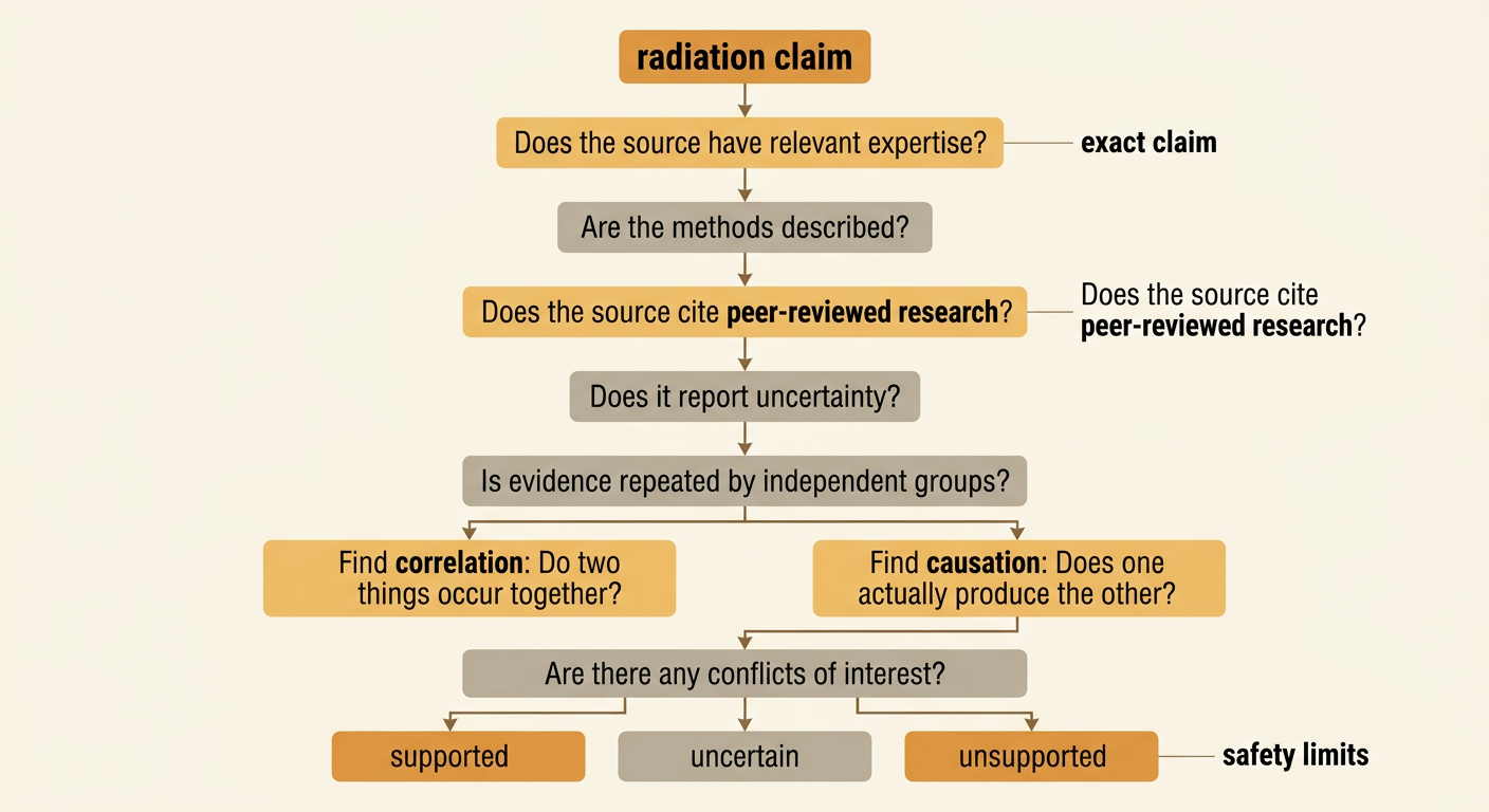
A scientific claim about radiation should be judged by the quality of its reasoning and evidence, not by how alarming or reassuring it sounds. First, identify the exact claim, the frequency range, the exposure level, and the stated effect. Then examine whether the source has relevant expertise, describes its methods, cites peer-reviewed research, and reports uncertainties. Strong conclusions usually depend on repeated findings from multiple independent sources. Check whether a study distinguishes correlation from causation and whether its test exposure resembles real-world exposure. Also look for sample size, comparison groups, possible conflicts of interest, and agreement with established evidence. For example, a post claiming that ordinary Wi-Fi exposure causes cancer is weak if it cites only personal stories. A stronger evaluation would compare measured radio-frequency exposure with safety limits and review controlled studies and large population studies, while acknowledging their limitations.
Evidence-Based Risk Conclusions
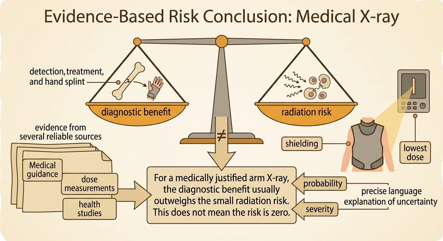
An evidence-based risk conclusion weighs both the probability and severity of harm against expected benefits. It should use precise claims, evidence from multiple reliable sources, and a clear explanation of uncertainty. It should also address reasonable counterclaims instead of ignoring them. Consider a medical X-ray for a possible broken arm. X-rays can damage cells because they are ionizing, so unnecessary exposure should be avoided. However, a properly performed image uses a controlled dose and may reveal a fracture that needs treatment. Evidence from medical organizations, radiation measurements, and health studies supports limiting exposure while preserving useful imaging. A strong conclusion might state that the diagnostic benefit usually outweighs the small radiation risk when the X-ray is medically justified. It should also acknowledge cumulative exposure and recommend shielding, appropriate equipment, and the lowest dose that produces an adequate image. New evidence could justify revising the conclusion.