
From DNA to Protein: Gene Expression and Genetic Privacy
Students model transcription and translation to explain how a DNA sequence determines protein structure, then consider how genetic information should be protected and used.
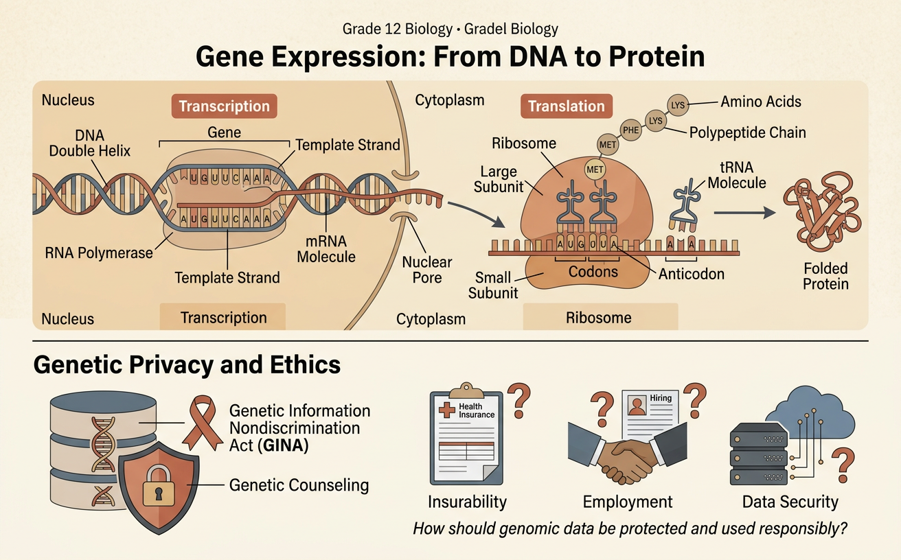
Illustrations are auto-generated and may be placeholders. They can be refreshed to match the narration.
The Central Dogma
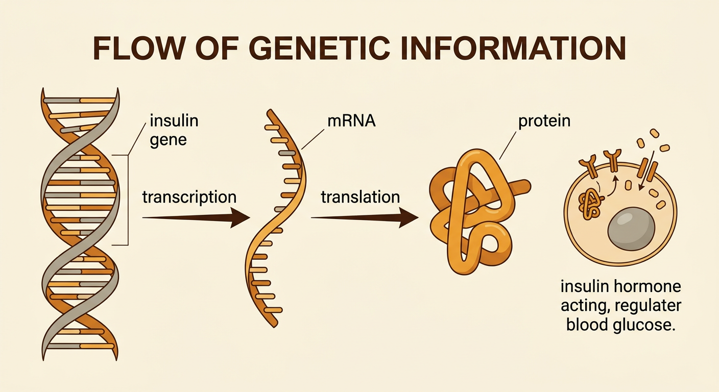
The central dogma describes the usual flow of genetic information: DNA is transcribed into RNA, and RNA is translated into protein. A gene is a DNA sequence that contains instructions for making a functional RNA or protein. For a protein-coding gene, the order of DNA bases determines the order of bases in messenger RNA, which determines the order of amino acids in a protein. The amino acid chain then folds into a three-dimensional shape that supports a particular function. For example, pancreatic cells express the insulin gene and produce insulin, a protein hormone that helps regulate blood glucose. Different cells contain mostly the same DNA, but they activate different genes. The central idea is that DNA affects cell structures and activities through gene expression, although environmental conditions and regulatory signals influence when and how strongly genes are expressed.
Transcription: DNA to mRNA
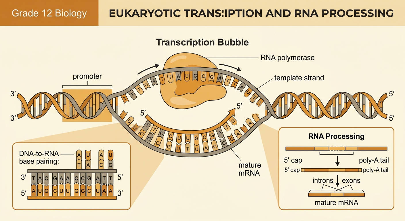
Transcription creates an RNA copy of a protein-coding gene. RNA polymerase binds near the gene's promoter, separates a short region of DNA, and uses one strand as the template. It moves along the template strand while adding complementary RNA nucleotides: DNA A pairs with RNA U, DNA T with RNA A, and C with G. Because the template is read from 3′ to 5′, the mRNA is built from 5′ to 3′. For example, the DNA template sequence 3′-TAC GAA CCG ATT-5′ produces the mRNA sequence 5′-AUG CUU GGC UAA-3′. In eukaryotic cells, the initial RNA transcript is processed before leaving the nucleus. A 5′ cap and poly-A tail are added, and introns are removed by splicing. The resulting mature mRNA carries the gene's coding information to a ribosome.
Translation: mRNA to Protein
Translation converts the nucleotide sequence of mRNA into an amino acid sequence. A ribosome binds the mRNA and reads it from 5′ to 3′ in groups of three bases called codons. Translation commonly begins at the start codon AUG, which codes for methionine. Transfer RNA molecules carry amino acids to the ribosome, and each tRNA anticodon pairs with a complementary mRNA codon. The ribosome forms peptide bonds between the amino acids and moves to the next codon. Using the mRNA sequence 5′-AUG CUU GGC UAA-3′, the ribosome adds methionine, leucine, and glycine. UAA is a stop codon, so no amino acid is added; instead, the short peptide is released. In actual cells, many proteins contain hundreds of amino acids. After translation, the chain folds and may be chemically modified before becoming functional.

Modeling a Sequence Change
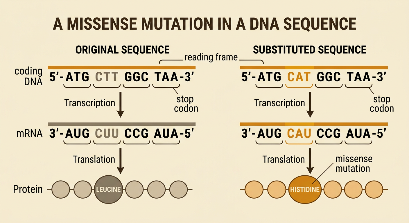
A mutation is a change in a DNA sequence, and its effect can be modeled by following the change through transcription and translation. Consider the coding DNA sequence 5′-ATG CTT GGC TAA-3′. It produces mRNA 5′-AUG CUU GGC UAA-3′ and the amino acid sequence methionine-leucine-glycine. If one DNA base changes so that CTT becomes CAT, the mRNA codon changes from CUU to CAU. The amino acid changes from leucine to histidine, creating a missense mutation. This substitution may alter protein folding or function, but its actual effect depends on the amino acid's location and chemical properties. Other substitutions may be silent because several codons can specify the same amino acid, or they may create an early stop codon. Insertions and deletions can shift the reading frame, changing every downstream codon unless the number of inserted or deleted bases is a multiple of three.
Proteins, Traits, and Cell Functions
Proteins perform essential jobs as enzymes, receptors, antibodies, transporters, signals, and structural materials. A protein's amino acid sequence influences how it folds, and its shape helps determine what it can do. A sequence change can therefore affect a cell and sometimes an observable trait. For example, a substitution in the HBB gene replaces glutamic acid with valine in the beta-globin protein. Under low-oxygen conditions, the altered hemoglobin molecules can stick together, distorting red blood cells into sickle shapes. These cells may block small blood vessels and carry oxygen less effectively. This evidence connects DNA structure to protein structure and then to cell function. However, genes do not act alone. Other genes, gene regulation, medical care, and environmental conditions can influence the severity of a trait. A scientifically accurate explanation should distinguish the direct molecular mechanism from broader factors that affect the final phenotype.

Genetic Information and Privacy
Genetic data can reveal health risks, biological relationships, and information shared with relatives, so it requires strong privacy protections. Imagine that a student submits DNA to a research study. Researchers may benefit society by identifying disease-related variants, but the student has interests in autonomy, confidentiality, and freedom from discrimination. Appropriate safeguards include informed consent, collecting only necessary data, secure storage, limited access, transparent sharing rules, and a clear process for withdrawing when possible. Removing a person's name reduces risk but may not guarantee anonymity because DNA is identifying and can sometimes be linked with other records. In the United States, the Genetic Information Nondiscrimination Act restricts many uses of genetic information by health insurers and employers, but it generally does not cover life, disability, or long-term care insurance. Civic decisions should balance scientific and public benefits with constitutional principles, human rights, equal treatment, and individual perspectives. People whose data are collected should have a meaningful voice in those decisions.
