
Teamwork Inside the Body: Organ Systems in Action
Students analyze what happens during exercise to explain how the respiratory, circulatory, muscular, and nervous systems work together.
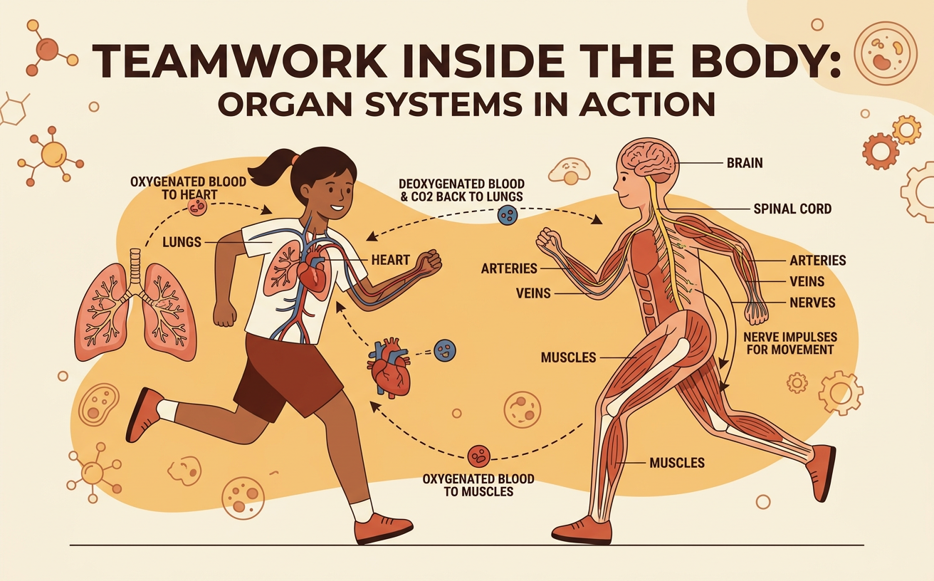
Illustrations are auto-generated and may be placeholders. They can be refreshed to match the narration.
The Body as a System
The human body is a system made of interacting subsystems called organ systems. Each organ system contains organs, and organs contain tissues made of groups of specialized cells. For example, muscle cells form muscle tissue, which helps make an organ such as the heart. During exercise, no organ system works alone. When a student runs across a soccer field, muscle cells need more oxygen and nutrients to release usable energy. The respiratory system brings oxygen into the lungs, the circulatory system transports it, the nervous system coordinates movement, and the muscular system produces force. A change in one subsystem affects the others. If breathing cannot supply enough oxygen, working muscles cannot continue at the same pace. This teamwork supports the body’s overall function and helps maintain stable internal conditions.
Changes During Exercise
Exercise increases the needs of active muscle cells. As a student begins jumping rope, breathing becomes faster and deeper, heart rate rises, and the skin may become warm and sweaty. These changes help deliver more oxygen and glucose to cells, remove carbon dioxide, and control body temperature. The nervous system detects information from inside and outside the body and helps coordinate these responses. Students can collect evidence by measuring pulse and breathing rate before and after one minute of exercise. For example, a pulse might rise from 75 beats per minute at rest to 125 beats per minute after exercise. One person’s measurements show what happened to that person at that time, but repeated measurements from several students provide stronger evidence of a general pattern.
Respiratory and Circulatory Teamwork
The respiratory and circulatory systems cooperate to exchange and transport gases. When a runner inhales, air travels through the airways to tiny lung sacs called alveoli. Oxygen moves across the thin alveolar walls into nearby capillaries, while carbon dioxide moves from the blood into the alveoli to be exhaled. The heart then pumps oxygen-rich blood through arteries toward working leg muscles. Oxygen moves from capillaries into muscle cells, where it supports the release of usable energy from food. Carbon dioxide produced by the cells enters the blood and returns through veins to the heart and then the lungs. During a fast run, quicker breathing increases air exchange, while a faster heartbeat moves blood more rapidly. Together, these systems connect the outside air to cells throughout the body.
Muscular and Nervous System Roles
The nervous system directs and adjusts movement, while the muscular system produces force. When a student decides to kick a ball, the brain sends electrical signals through the spinal cord and motor neurons to leg muscles. At connections between neurons and muscle fibers, chemical signals trigger the muscle fibers to contract. Muscles often work in opposing pairs. To straighten the knee, the quadriceps contract while the hamstrings relax; bending the knee reverses much of this pattern. Sensory receptors in the eyes, skin, joints, and muscles send information back to the brain about the ball’s location and the leg’s position. The brain uses this feedback to correct the movement. Muscle cells also depend on oxygen and glucose delivered by blood, showing that nervous and muscular actions rely on other body systems.
Modeling System Interactions
A model can simplify how organ systems interact during exercise. Begin with active muscle cells at the center. Draw an arrow from the respiratory system to the circulatory system to represent oxygen entering the blood. Draw another arrow from the circulatory system to the muscles to represent delivery of oxygen and glucose. Return arrows can show carbon dioxide moving from muscles to blood and then to the lungs. Add arrows from the nervous system to breathing muscles, the heart, and skeletal muscles to show coordination. For example, during a bicycle ride, faster nerve signals to leg muscles support repeated contractions while increased breathing and blood flow support the cells. Models reveal connections and help predict effects, but they have limitations. An arrow diagram does not show every organ, cell, chemical reaction, or change over time.
Evidence-Based Explanation
A strong scientific explanation includes a claim, specific evidence, and reasoning that connects the evidence to the claim. A student might claim that the respiratory, circulatory, muscular, and nervous systems interact during exercise. Evidence could include a text stating that oxygen crosses from alveoli into blood, a diagram showing blood flow to muscles, and class data showing that average heart and breathing rates increased after running. The reasoning should explain that contracting muscle cells need increased delivery of oxygen and removal of carbon dioxide, so breathing and circulation change together under nervous system coordination. Sources also have strengths and limitations. Class data are direct but may include counting errors or a small sample. A textbook is reviewed and detailed but does not measure the class. Using several sources makes the argument stronger because different evidence supports different parts of the explanation.